Urogenital Mycobacterial Infections
This article primarily focuses on genitourinary tuberculosis, with a brief discussion at the end regarding the side effects and management of intravesical BCG treatment.
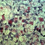
Despite advances in chemotherapy, the incidence of genitourinary tuberculosis, a slowly progressive and destructive disease, has not changed significantly. Symptoms such as frequency, dysuria, and hematuria are the most common presenting features. While findings on intravenous pyelography can aid in diagnosis, a definitive diagnosis typically relies on positive urine or pus cultures for pathogenic mycobacteria or histopathological evidence of tuberculosis, or both.
The primary treatment for genitourinary tuberculosis is medical, as surgical treatment is not considered an alternative. However, surgical intervention may be used as adjuvant therapy in certain cases.
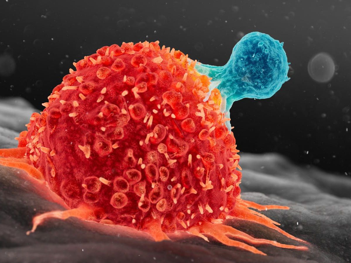
Intravesical therapy with BCG has shown to be more effective than most chemotherapeutic agents in the treatment and prophylaxis of superficial bladder tumors. Although instillations with BCG tend to elicit more local and systemic reactions compared to intravesical chemotherapy, in most cases, additional treatment is not necessary.
Adolescence and Age
The ratio of male to female in genitourinary tuberculosis ranges between 1.5-3, with the most frequent occurrences observed between the ages of 20 and 50. Patients under the age of twenty-five accounted for 40% of total genitourinary tuberculosis cases in 1956, but this proportion decreased to 12.6% between 1970 and 1979. Kidney tuberculosis was common in children before the era of chemotherapy, with 28.6% of patients dying from kidney tuberculosis between 1928 and 1949 being children. However, it is currently very rare in the pediatric age group, constituting less than 3% of all tuberculosis cases.
Etiology and Pathogenesis
Mycobacterium tuberculosis is the most commonly identified pathogen, although cases attributed to M. bovis may arise, particularly in underdeveloped countries. Genitourinary tuberculosis is almost always secondary and occurs through organisms transmitted via the bloodstream. The primary focus is typically in the lungs. Genitourinary tuberculosis, apart from rare instances of sepsis, emerges approximately 7-10 years after pulmonary tuberculosis; however, this duration can vary from 2 to 40 years. The pathological progression of the disease involves a combination of caseation, cavitation, and fibrosis, depending on the organism’s virulence and the patient’s resistance.
Clinical Findings
Clinical manifestations suggestive of genitourinary tuberculosis include:
- Chronic cystitis that does not respond to treatment (60-95%). Genitourinary tuberculosis should be considered even in cases of recurrent cystitis that respond well to antibiotics.
- Macroscopic or microscopic hematuria
- Presence of sterile pyuria upon examination of urine sediment
- Presence of a painless or painful, large, firm epididymis, with thickening or bead-like palpable vas deferens
- A scrotal sinus, almost pathognomonic, with chronic discharge
- Palpable thickening of one or both seminal vesicles or the prostate upon rectal examination, which may be nodular or firm
- Flank pain can occur in 10-64% of cases. The rate of hypertension associated with genitourinary tuberculosis can vary from 0-23.2%. The classic triad of weakness, weight loss, and anorexia is not always present in the early stages of the disease.
Investigations
Tuberculin Test:
Positive in 85-95% of cases. A conversion from negative to positive is more significant (9,16).
Urinalysis:
Abacterial pyuria observed in 24-84% of cases (9,16). Secondary infections caused by non-specific organisms are found in 15-25% of cases. Microscopic hematuria is seen in 28-73% of cases. Urine pH is almost always acidic (1,9,11).
Demonstration and isolation of TB bacilli in urine are crucial for definitive diagnosis. For this, morning urine samples are collected for three consecutive days. Collecting urine for 24 hours is unnecessary.
Ziehl-Neelsen Staining (Direct AFB): Provides positive results in 48-60% of cases. False positives may occur due to non-pathogenic, non-tuberculous mycobacteria (10,12). Löwenstein-Jensen cultures yield growth rates of 49-90%. Results are typically obtained within 3-4 weeks, but no later than two months.
BACTEC TB Radiometric Culture: Results can be obtained in 1-2 weeks, with higher sensitivity than conventional methods. The gen-probe amplified M. tuberculosis direct test (MDT) provides results in four hours, with sensitivity and specificity close to 100% (6,17).
Radiography
30-80% of kidney TB patients have a history or previous lung TB detected by X-ray.
On direct abdominal X-ray, enlargement of the kidney shadow and calcifications related to the genitourinary organs (20-60%) may be observed.
Intravenous pyelography reveals abnormal findings in 58-95% of cases, including ulcerative appearance in the calyces, narrowing, obstruction, and sometimes absence of calyces (auto-amputated calyces), dilatation (hydronephrosis) due to ureteral stricture, fibrosis, and flattening, cavities (abscess cavities) connected to isolated or calyceal systems, kidney damage due to complete ureteral obstruction with no filtration (auto-nephrectomy), and later-stage contracted, small, irregular bladder. Simultaneous occurrence of numerous abnormalities in the upper and lower urinary systems is a valuable radiological finding for kidney TB.
Patients with positive urine or pus cultures for pathogenic mycobacteria, or histopathological evidence of TB, or both, are considered to have “definite TB.” If positive urine cultures for mycobacteria cannot be obtained, patients with three or more of the following findings on urological imaging or endoscopy may be considered to have “probable TB”: ureteral strictures, renal calcifications, renal infundibular strictures or closed calyces, “golf hole” ureteral orifices, small-capacity bladder, nodular or thickened epididymis, and cold psoas abscess. In a study from India, probable TB diagnosis was possible in 30% of patients, all of whom responded positively to empirical treatment.
Differential Diagnosis in Genitourinary Tuberculosis
Differential diagnosis of genitourinary TB includes chronic nonspecific urinary infection, acute or chronic nonspecific epididymitis, abacterial (sterile) cystitis, interstitial cystitis, multiple small renal cysts and nephrocalcinosis, necrotizing papillitis, medullary sponge kidney, disseminated coccidioidomycosis, urinary schistosomiasis, and invasive transitional cell carcinoma (12).
Treatment
The primary treatment for genitourinary TB is medical therapy. Surgical intervention is not an alternative but may be used adjunctively when necessary. While genitourinary TB treatment used to be administered for two years until the 1970s, shorter treatment regimens have become more prevalent in recent years. The reasons for the good response of genitourinary TB to short-term chemotherapy are as follows: in kidney TB, there are fewer bacilli present in the environment compared to pulmonary TB, and they are intermittently excreted in the urine; the urine concentrations of isoniazid, rifampicin, pyrazinamide, and streptomycin are high; isoniazid and rifampicin easily penetrate into renal cavities at high concentrations; all these drugs reach the kidney, ureter, bladder, and prostate at adequate concentrations. The role of steroids in the treatment of genitourinary TB is limited: 1- Prednisolone may be used in a dose of 3×20 mg for four weeks in combination with antituberculous drugs in acute TB cystitis. 2- Prednisolone in the same dose may be given for three weeks in the treatment of ureteral strictures unresponsive to chemotherapy (1,2,7).
Genitourinary TB is a slowly progressive and destructive disease. Tissue damage can accelerate further with the development of obstructive strictures after the initiation of medical treatment. Renal damage due to TB can occur unpredictably, and despite advancements in chemotherapy, some kidneys are inevitably lost. Complicated surgeries are futile in patients with GFR < 15 ml/min, cortical thickness < 5 mm, and proximal obstructive lesions (18).
Follow-Up
Patients should be seen at 3, 6, and 12 months after completing chemotherapy. Sequential three-day morning urine and IVP are requested at each follow-up. If radiographic results are stable and urine is sterile, and there are no ureteral strictures or renal calcifications, the patient may be discharged from follow-up, with instructions to return if they experience recurrent urinary symptoms.
Surgical Treatment
Surgical intervention should ideally be performed at least 15 days after initiating drug therapy, preferably 4-6 weeks later. Surgical procedures may involve tissue removal (nephrectomy, ureterectomy, partial nephrectomy, epididymectomy ± orchiectomy) or reconstructive surgery (ureteral strictures, augmentation cystoplasty, urinary diversions).
According to Gow et al., indications for nephrectomy include a non-functioning kidney (with or without calcification), widespread disease involving the entire kidney in association with hypertension and pelviureteric obstruction, and concomitant kidney cancer.
According to Lattimer et al., indications for nephrectomy include severe pain, significant urinary bleeding, urinary bacterial sepsis, suspicion of renal malignancy, and patients resistant or intolerant to chemotherapy. These researchers oppose nephrectomy solely due to a non-functioning kidney. Nephrectomy rates in cases of renal TB can reach up to 60% .
Partial nephrectomy is indicated for calcified local lesions that do not respond to six weeks of treatment and are progressively increasing in size. If there is no calcification, there is no indication for partial nephrectomy.
Ureteral strictures, a complication that can lead to kidney loss in up to 20-40.7% of patients, are most commonly observed at the ureterovesical junction (9,10,15). Treatment of ureteral strictures is carried out progressively from simple to complex methods, including medical treatment (chemotherapy ± steroids), ureteral dilation (with balloon dilators), and surgical treatment (ureteroneocystostomy, Boari flap, psoas hitch).
Medical treatment of ureteral strictures involves weekly IVPs for 25 minutes when chemotherapy is initiated in the presence of ureteral obstruction. If there is no improvement or worsening after three weeks, prednisolone 3×20 mg/day is added to the antituberculous drugs. The same procedure is repeated for another three weeks with weekly IVPs. If there is no improvement or worsening after six weeks, ureteral dilation is performed, and if unsuccessful, surgical reimplantation is considered.
Augmentation cystoplasty is performed to increase bladder capacity in patients with unbearable urgency, frequency, and hematuria, with a creatinine clearance above 15 ml/min. It is contraindicated in enuresis, incontinence, and psychiatric disorders.
Urinary diversion in tuberculosis:
Three indications for permanent urinary diversions include psychiatric disorders or mental retardation, enuresis, and intolerable diurnal symptoms associated with incontinence resistant to chemotherapy or bladder dilation. Ileal or colonic conduits are preferred for permanent diversions.