Introduction
Bacteria of the Pseudomonas genus belong to the family Pseudomonadaceae. Most of these bacteria are abundantly found in nature, particularly in soil and water. Some species are pathogens for humans, animals, and plants. Within this genus, some species are oxidase-positive while others are oxidase-negative. They are bacteria that oxidize glucose but do not ferment it. All species are catalase-positive, Gram-negative, aerobic, rod-shaped bacteria capable of movement with polar flagella.
There are several features that make Pseudomonas species important for food. Some species exhibit proteolytic and lipolytic activities. Due to their aerobic nature, they can rapidly develop on the surface of foods, leading to the formation of oxidized products and mucous substances. They have the ability to synthesize the growth factors and vitamins necessary for their own growth. There are psychrophilic, mesophilic, or psychrotrophic species within the genus.
Especially, when stored in cold conditions, such as milk, meat, eggs, and seafood, they are the primary agents of spoilage. They can be easily inhibited by heat and radiation. They cannot grow when there is no oxygen and above 42°C. They have weak resistance to desiccation. Some species, such as Pseudomonas fluorescens, produce greenish pigments, while Pseudomonas nigrificans produces black and other species produce brown pigments on certain foods.
Members of this genus are clinically important because they are resistant to many antibiotics and can only survive under conditions tolerated by a few bacteria.
Various sources indicate that Pseudomonas bacteria mixed with milk can rapidly proliferate, leading to various fermentations and breakdowns, resulting in changes in the color, odor, structure, and consistency of milk. Pseudomonas in milk not only deteriorates the freshness of the milk but sometimes also causes various infections. Especially, Pseudomonas aeruginosa, known as an opportunistic pathogen, can lead to diseases and outbreaks when not careful about the cleanliness of milk containers, particularly in children aged 0-3 years, where milk consumption is high.
Characteristics of Pseudomonas aeruginosa
Historical Background
P. aeruginosa was first identified in 1850 by Sedillot as an agent causing a blue color change in surgical wound dressings. Initially known as Bacillus pyocyaneus and later as Pseudomonas pyocyanea, the isolation of pyocyanin was performed by Lucke in 1862. However, it was not until Gessard’s classic studies in 1882 that this organism was isolated in pure culture. Hitschman and Kreibich in 1897, Frenkel in 1917, and Osler in 1925 described it as a pathogenic bacterium. In 1926, Dooren de Jong at the University of California classified Pseudomonas species based on phenotypic traits, relying on the use of various organic compounds as carbon and energy sources. In 1966, Buchanon, Holt, and Lessel classified Pseudomonas species based on phenotypic characteristics. Subsequently, DNA hybridization studies began. In 1973, Palleroni and colleagues expanded nucleic acid hybridization studies to classify Pseudomonas into 5 groups based on rRNA homologies. In recent years, the classification of this genus has been reorganized in light of new research.
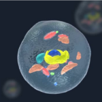
Morphological Characteristics
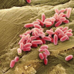
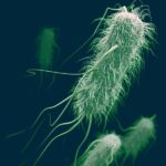
Although their lengths vary greatly, Pseudomonas aeruginosa typically appears as non-spore-forming, capsule-free, rod-shaped aerobic bacteria, ranging from 1.5 to 3 µm in length, sometimes seen in pairs or short chains. They often possess one or occasionally two to three flagella at one end, making them highly motile. They stain easily and are Gram-negative. Deformed shapes, motionless and pigmentless forms, and those reproducing in the R (rough) type have been reported in cultures that have been left for a long time or in environments containing antiseptic agents.
Cultural and Biochemical Characteristics
Pseudomonas aeruginosa thrives in suitable media at optimal temperatures of 30-37°C and slightly alkaline conditions. Its ability to grow at 41°C is an important characteristic, and being able to grow consecutively for 3 passages at 42°C distinguishes it from P. fluorescens. Despite being aerobic, it can also be found among anaerobic species due to its denitrification properties. In liquid media, it shows dense and homogeneous growth on the surface, with distinguishable green-blue pigment just below the surface film. Cultures left for an extended period become alkaline, causing bacteria to dissolve with lytic enzymes, clearing the liquid medium. They also grow similarly in peptone water.
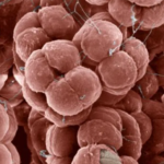
On solid media, P. aeruginosa forms 3 types of colonies. Type 1 colonies, often isolated from clinical samples, are round, matte-surfaced, slightly raised in the middle, flat, white-colored colonies with a green-blue pigment spread throughout the medium, measuring 2-3 mm in diameter. Type 2 colonies, mostly isolated from natural sources, resemble smaller, raised, convex, irregular coliform colonies. Type 3 colonies are R colonies formed by bacteria with extracellular alginate secretion, giving them a mucoid appearance. They produce a characteristic fruity odor due to the production of tryptophan 2-aminoacetophenone and emit a grape or trimethylamine odor when the lid of the Petri dish is opened.
Some biochemical characteristics of Pseudomonas aeruginosa include:
- They cause hemolysis on blood agar. Clinical isolates growing on blood agar are often beta-hemolytic.
- They hydrolyze gelatin and coagulate plasma.
- They oxidize glucose via oxidative pathways, producing acid (gluconate), but cannot utilize lactose and sucrose.
- They are distinguished from members of the Enterobacteriaceae family by being oxidase-positive.
- They produce ammonia by deaminating acetamine.
- They do not affect starch.
- They are catalase and citrate positive.
- They produce L-arginine dihydrolase but not lysine decarboxylase or ornithine decarboxylase.
- They do not produce indole or H2S.
- They are methyl red and Voges-Proskauer negative.
- They reduce nitrate to nitrite.
- They reduce tetrazolium salts and selenite.
- They are resistant to KCN.
- Pseudomonas aeruginosa, unlike P. fluorescens, decolorizes methylene blue and prontosil.
Most Pseudomonas strains produce pigments in culture media, with pigment formation dependent on culture conditions. This trait may be lost through mutation or multiple pigment formations may occur simultaneously. These pigments do not form in anaerobic conditions and are better formed at room temperature.
Fluorescent pigments are characteristic of Pseudomonas species exhibiting fluorescence properties (P. aeruginosa, P. putida, P. fluorescens, P. chlororaphis, P. syringae, P. cichori, P. flavescens). These pigments are siderophores produced abundantly in low iron concentrations in culture media. King B medium is used to stimulate the production of fluorescent pigments. Fluorescein is a fluorescent pigment insoluble in chloroform but soluble in water, appearing yellowish. UV light may be needed to observe this pigment. 70% of clinical isolates produce this pigment on King B agar. Piovverdin, a fluorescent pigment, was isolated from a reference Pseudomonas strain (PAO1).
Historical Insight
The presence of extracellular secretions with proteolytic enzymes, lethal exotoxins, and enterotoxins, along with its opportunistic pathogenic nature, leads to the development of various diseases. Pseudomonas species can be isolated from conditions such as urinary tract infections, ocular infections, external otitis, otitis media, burn and wound infections, meningitis, bronchitis, bronchopneumonia, septicemia, osteomyelitis, and pseudomembranous colitis.
Epidemic Impact
Reports have indicated that P. aeruginosa has caused epidemic diarrhea resulting in the death of newborns. In hospital settings, especially in immunocompromised patients, it can pose a life-threatening risk by causing diarrhea.
Gastroenteritis and Milk Contamination
A study conducted in 1946 reported cases of acute epidermal gastroenteritis resulting from the ingestion of milk contaminated with P. aeruginosa. Symptoms included diarrhea, cramps, nausea, and vomiting in 409 cases, with more severe symptoms observed in infants and children, resulting in 9 deaths. It was suggested that oral intake of P. aeruginosa in a dose of 10^6 could cause gastroenteritis.
Presence and Significance of Pseudomonas Aeruginosa in Raw Milk
The contamination of milk with microorganisms depends on the conditions during milk production. Factors such as the prolonged storage of milk at low temperatures on farms, cleanliness and temperature of tanks brought to the facilities, determine the bacteriological characteristics of milk. Pseudomonas aeruginosa, being a bacterium commonly found in soil, water, and animal skin, can contaminate milk if hygienic conditions are not maintained during milking. Pseudomonas species can colonize milk equipment and hoseheads. In cases where iodine-based disinfectants are used at low levels, Pseudomonas can produce a sticky substance called glyoxal. This substance allows the organism to adhere better to surfaces, thereby forming colonies on equipment surfaces and increasing their resistance to antimicrobial agents, phagocytes, and surfactants. Eliminating Pseudomonas contaminated water and hoses is quite challenging due to the strong resistance mechanism of the bacteria.
In milk stored in cold conditions, the presence of highly heat-resistant esterases (proteases-lipases) secreted by Pseudomonas group bacteria and the reactivation of these enzymes in milk products after pasteurization can lead to product-related issues. For example, the breakdown of triglycerides in milk, cheese, cream, and butter results in souring. These enzymes maintain their stability during UHT processing, causing enzymatic errors called sweet curdling in UHT milk without acidity development and precipitating proteins at the bottom of the container.